

Versions
- 2025-07-01 (2)
- 2025-04-23 (1)
Keywords
Image quality
Inferior alveolar duct
How to Cite
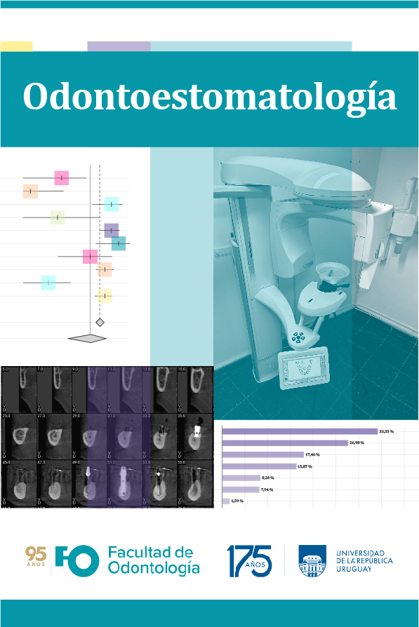
Abstract
Objective. To evaluate the influence of x-ray tube current and voltage on the localization of the alveolar duct by linear tomography. Material and Methods. The sample consisted of 24 hemi-mandibles that were scanned by linear tomography equipment using different combinations of Kv and mA. The images obtained were processed using ImagJ software, obtaining coronal slices of the images, which were then observed by five observers. Diagnostic accuracy was compared by the area under the curve (Az) through ROC curves (receiver operating characteristic curve), using ROCKIT 1.1B software. The intra- and inter-observer Kappa index was 0.79 and 0.71, respectively. Results. Using low values of Kv (60 Kv), higher Az (0.912) was achieved. When mA was varied, the highest Az value was for 2 mA (0.911). Conclusion. It is recommended to use low values of RX tube current and voltage.
References
Muñoz G, Dias FJ, Weber B, Betancourt P, Borie E. Anatomic Relationships of Mandibular Canal. A Cone Beam CT Study. Int J Morphol. 2017;35(4):1243–8. Disponible en: https://www.scielo.cl/scielo.php?pid=S0717-95022017000401243&script=sci_abstract
Domínguez Mejía J, Ruge Jiménez O, Aguilar Méndez G, Ñañez López Ó, Oliveros Torres G. Análisis de la posición y trayectoria del conducto alveolar inferior (CAI) en tomografía volumétrica computarizada (TC Cone Beam -TCCB). Rev Fac Odontol Univ Antioq 2010 Dec;22(1): 12-22. Disponible en: http://www.scielo.org.co/scielo.php?script=sci_arttext&pid=S0121-246X2010000200003
Canelo Martínez M. Uso de la tomografía computarizada de haz cónico en la detección de variantes anatómicas del conducto dentario inferior. Santo Domingo:Universidad Iberoamericana (UNIBE); 2021. Disponible en: https://repositorio.unibe.edu.do/jspui/handle/123456789/771
Beltrán Silva JA, Abanto Silva LE, Meneses López A. Disposición del conducto dentario inferior en el cuerpo mandibular: Estudio anatómico y tomográfico. Acta odontol. venez. 2007 Sep;45( 3 ): 421-425. Disponible en: http://ve.scielo.org/scielo.php?script=sci_arttext&pid=S0001-63652007000300018
Kamrun N, Tetsumura A, Nomura Y, Yamaguchi S, Baba O, Nakamura S, et al. Visualization of the superior and inferior borders of the mandibular canal: a comparative study using dig- ital panoramic radiographs and cross-sectional computed to- mography images. Oral Surg Oral Med Oral Pathol Oral Radiol 2013; 115: 550–7. Disponible en: https://pubmed.ncbi.nlm.nih.gov/23522648/
Solis Vargas LD. Ortopantomografía (OPG) vr. Tomografía Computada (CT) en Imágenes Odontológicas Dentales. Revista Ciencia y Salud Integrando Conocimientos. 2023;7(1):43–58. Disponible en: https://revistacienciaysalud.ac.cr/ojs/index.php/cienciaysalud/article/view/576
Buzzi A.E., Suárez M.V. Linear Tomography: Birth, glory and decline of a method. Rev. Argent. Radiol.2013; 77(3):236-244.
Flores Choquehuanca D, Moya Chávez LA. Tomografía Odontológica. Rev. Act. Clin. Med. 2013 Set. Disponible en: http://www.revistasbolivianas.ciencia.bo/scielo.php?script=sci_arttext&pid=S2304-37682013001100008&lng=pt&nrm=iso
Beltrán Silva JA, Meneses López A, Ventura Ponce HR. Tomografía espiral convencional para implantes dentales: grado de magnificación. Rev Estomatol Hered. 2014;13(2–1). Disponible en: https://revistas.upch.edu.pe/index.php/REH/article/view/2048
Jaju PP, Jaju SP. Cone-beam computed tomography: Time to move from ALARA to ALADA. Imaging Sci Dent 2015. Disponible en: https://pubmed.ncbi.nlm.nih.gov/26730375/
Widmann G, Schönthaler H, Tartarotti A, Degenhart G, Hörmann R, Feuchtner G, et al. As low as diagnostically acceptable dose imaging in maxillofacial trauma: a reference quality approach. Dentomaxillofac Radiol 2023. Disponible en: https://pubmed.ncbi.nlm.nih.gov/36688730/
Pauwels R, Silkosessak O, Jacobs R, Bogaerts R, Bosmans H, Panmekiate S. A pragmatic approach to determine the optimal Kv p in cone beam CT: balancing contrast-to-noise ratio and ra- diation dose. Dentomaxillofac Radiol 2014; 43: 20140059.Disponible en: https://doi.org/10.1259/dmfr.20140059
Svenson B, Welander U, Anneroth G, Soderfeldt B. Exposure parameters and their effects on diagnostic accuracy. Oral Surg Oral Med Oral Pathol 1994; 78: 544–50. Disponible en: https://pubmed.ncbi.nlm.nih.gov/7800386/
Neves FS, de Camargo T, de Azevedo Vaz SL, Campos PS, Boscolo FN. Influence of cone-beam computed tomography milliamperage settings on image quality of the mandibular third molar region. Oral Radiol 2014; 30: 27–31. Disponible en: https://pubmed.ncbi.nlm.nih.gov/25265127/
Oliveira ML, Freitas DQ, Ambrosano GM, Haiter-Neto F. In- fluence of exposure factors on the variability of CBCT voxel values: a phantom study. Dentomaxillofac Radiol 2014; 43: 20140128. doi: https://doi.org/10.1259/dmfr.20140128
Jasa GR, Shimizu M, Okamura K, Tokumori K, Takeshita Y, Weerawanich W, et al. Effects of exposure parameters and slice thickness on detecting clear and unclear mandibular canals using cone beam CT. Dentomaxillofac Radiol 2017. Disponible en: https://www.ncbi.nlm.nih.gov/pmc/articles/PMC5595001/
Shimizu M, Okamura K, Kise Y, Takeshita Y, Furuhashi H, Weerawanich W, Moriyama M , Ohyama Y, Furukawa S, Nakamura S and Yoshiura S. Effectiveness of imaging modalities for screening IgG4-related dacryoadenitis and sialadenitis (Mikulicz’s disease) and for differentiating it from Sjögren’s syndrome (SS), with an emphasis on sonography. Arthritis Res Ther 2015;17(1):223. Disponible en: https://www.ncbi.nlm.nih.gov/pmc/articles/PMC4546818/
Hanley JA. Receiver operating characteristic (ROC) methodology: the state of the art. Crit Rev Diagn Imaging. 1989;29(3):307-35. Disponible en: https://pubmed.ncbi.nlm.nih.gov/2667567/
Lindh C, Petersson A, Klinge B. Visualisation of the mandibular canal by different radiographic techniques. Clin Oral Implants Res. 1992;3(2):90–7. Disponible en: http://dx.doi.org/10.1034/j.1600-0501.1992.030207.
Ludlow JB, Timothy R, Walker C, Hunter R, Benavides E, Samuelson DB, et al. Dosis efectiva de CBCT dental: un metaanálisis de datos publicados y datos adicionales para nueve unidades de CBCT. Dentomaxillofac Radiol [Internet]. 2015;44(1):20140197. Disponible en: http://dx.doi.org/10.1259/dmfr.20140197
Pauwels R, Araki K, Siewerdsen JH, Thongvigitmanee SS. Technical aspects of dental CBCT: state of the art. Dentomax-illofac Radiol 2015; 44: 20140224. Disponible en: https://doi.org/10.1259/dmfr.20140224
Panmekiate S, Apinhasmit W, Peterson A. Effect of electric po-tential and current on mandibular linear measurements in conebeam CT. Dentomaxillofac Radiol 2012; 41: 578–82. Disponible en: https://doi.org/10.1259/dmfr/51664704.

This work is licensed under a Creative Commons Attribution-NonCommercial 4.0 International License.
Copyright (c) 2025 Juan Manuel González Mollo, Carolina Riera Laiño, Rocio Mott Gutierrez, Flavio Echeverria Lopez, Gainer Jasa Andrade